split-banner-image

LAURA’S STORY

We spoke with Laura about her shock diagnosis, her decision to participate in the Breast MRI Study and her advice to other young women who have also received a diagnosis.

Being Diagnosed With Breast Cancer At 31

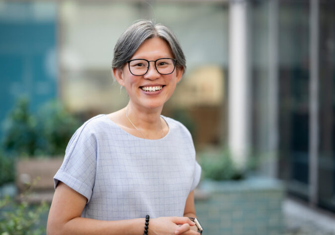
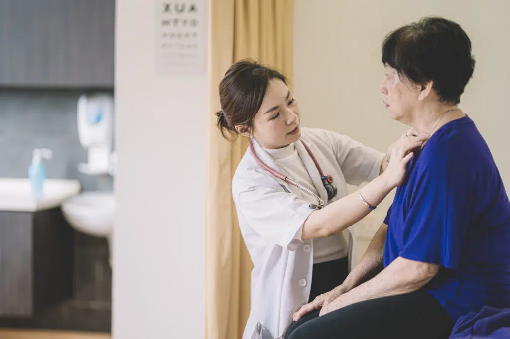
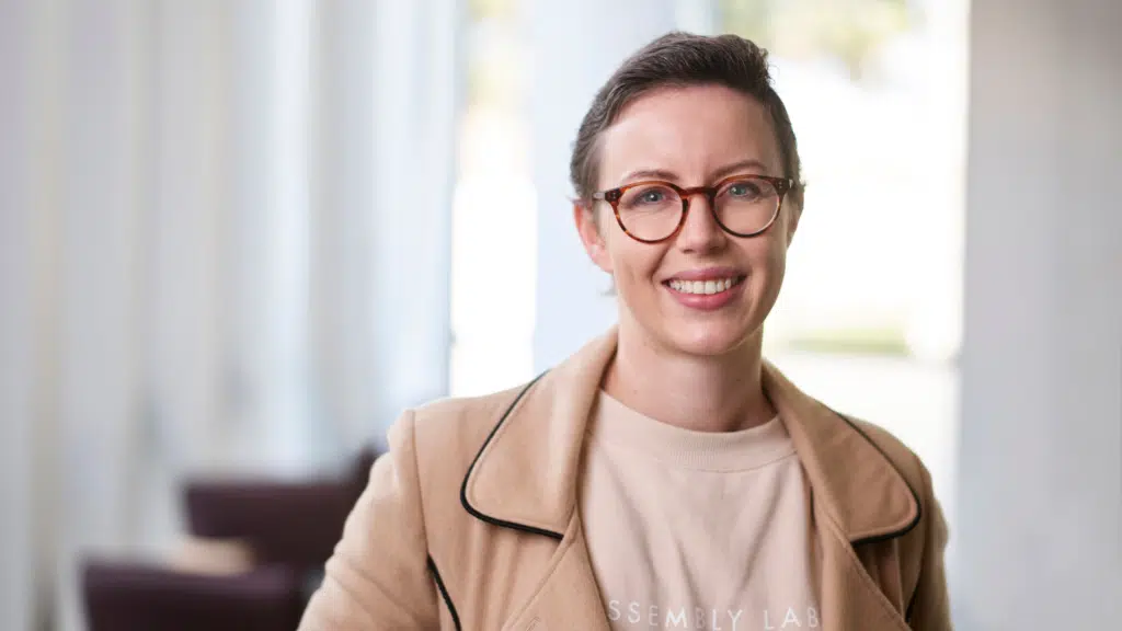
Laura McCambridge is a project manager coordinating clinical trials in stroke and dementia at the Florey Institute of Neuroscience and Mental Health in Melbourne where she lives with her partner and their golden retriever puppy.

At 31, Laura found a lump in her breast and was shortly after diagnosed with breast cancer.

We spoke with Laura about her shock diagnosis, her decision to participate in the breast MRI evaluation study and her advice to other young women who have also received a diagnosis.

“My name is Laura. I’m 31 and from New Zealand originally but I’ve been living in Melbourne for the past 2.5 years,” she said.

“My connection with breast cancer is that in September of last year I found a lump in my breast and I went to the G.P.”

“He thought everything looked fine but sent me off for an ultrasound just in case.”

“The radiologists thought that it was probably fine as well but sent me off for a biopsy just to be safe again, and then when the pathology came back it was cancerous cells unfortunately.”

“So, since then, I’ve been walking through my breast cancer journey.”

“So, I had a lumpectomy and a sentinel node biopsy, and I was lucky that my love nodes were negative, but I still needed to have four cycles of chemo and then I was just about to get started with my radiation journey, but my gene markers came back that I had a mutation in the CHEK2 gene.”

“So, my surgeon thought that it would be best to go for a bilateral mastectomy, and I had that in February of this year, so now I’m kind of finished with my active treatment, which is exciting and just continuing with hormone therapy for 5-10 years now.”

Listen to the Podcast

At 31, Laura found a lump in her breast and was shortly after diagnosed with breast cancer. We spoke with Laura about her shock diagnosis, her decision to participate in the Breast MRI Evaluation Study and her advice to other young women who have also received a diagnosis.

Participating In The Breast MRI Evaluation Study

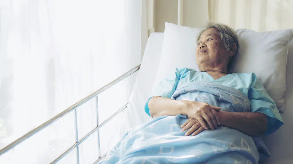
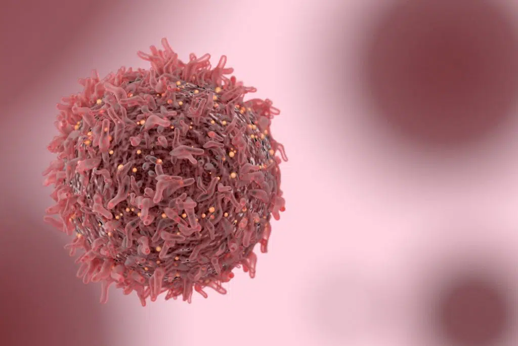
Laura was diagnosed with invasive breast carcinoma.

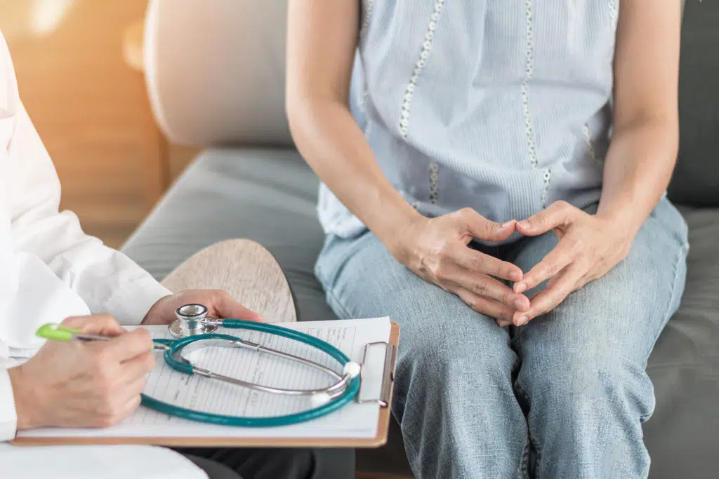
She underwent months of treatment, including a lumpectomy, four cycles of chemotherapy, a mastectomy and hormone therapy.

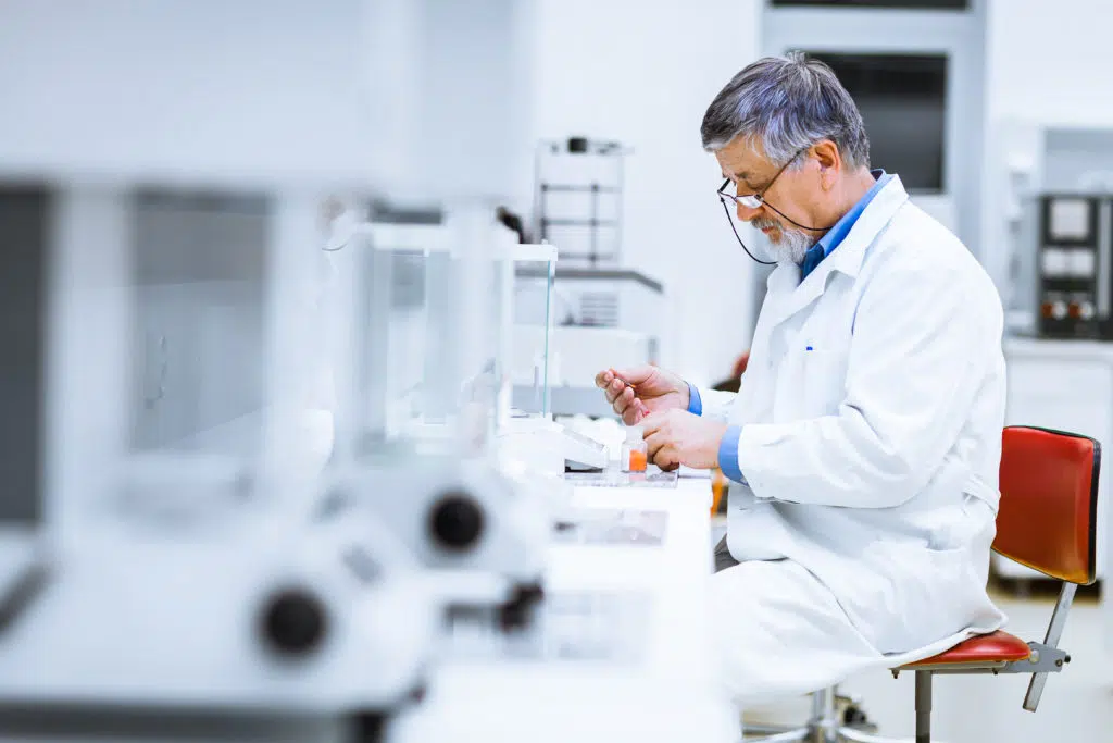
The Breast MRI Evaluation Study aims to find out the best way to use breast Magnetic Resonance Imaging (MRI) and if it will improve treatment options and patient outcomes, in women recently diagnosed with breast cancer.

Laura was offered a place in this study and didn’t hesitate to join.

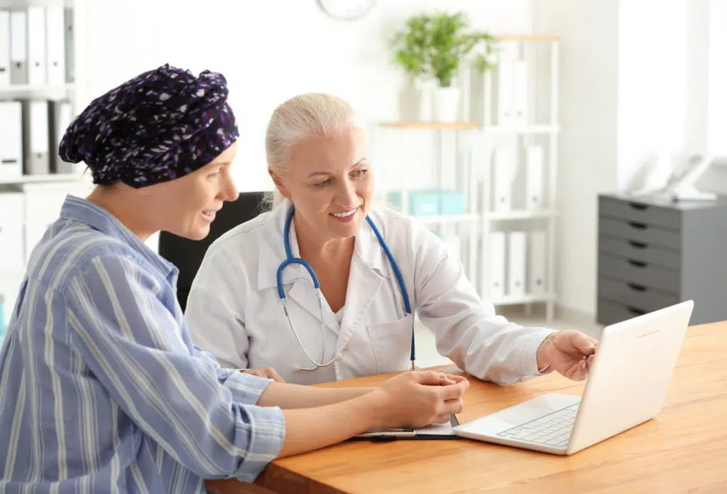
“I heard about the Breast MRI Evaluation Study by talking to my surgeon, and I am, I work in research myself, so I was interested in being a participant seeing the other side of research.”

“So when I was meeting with my surgeon, I asked her are there any research studies available through this hospital and she put me in touch with the research coordinator of the Breast MRI Study.”

“I took it from there with her and there was a really positive experience being able to form a relationship with the research coordinator. We’re on really good terms texting each other, and she was checking in not just to follow up with me for follow up assessments but just to see how I was which was really nice.”

The Benefits Of Participating In A Clinical Trial

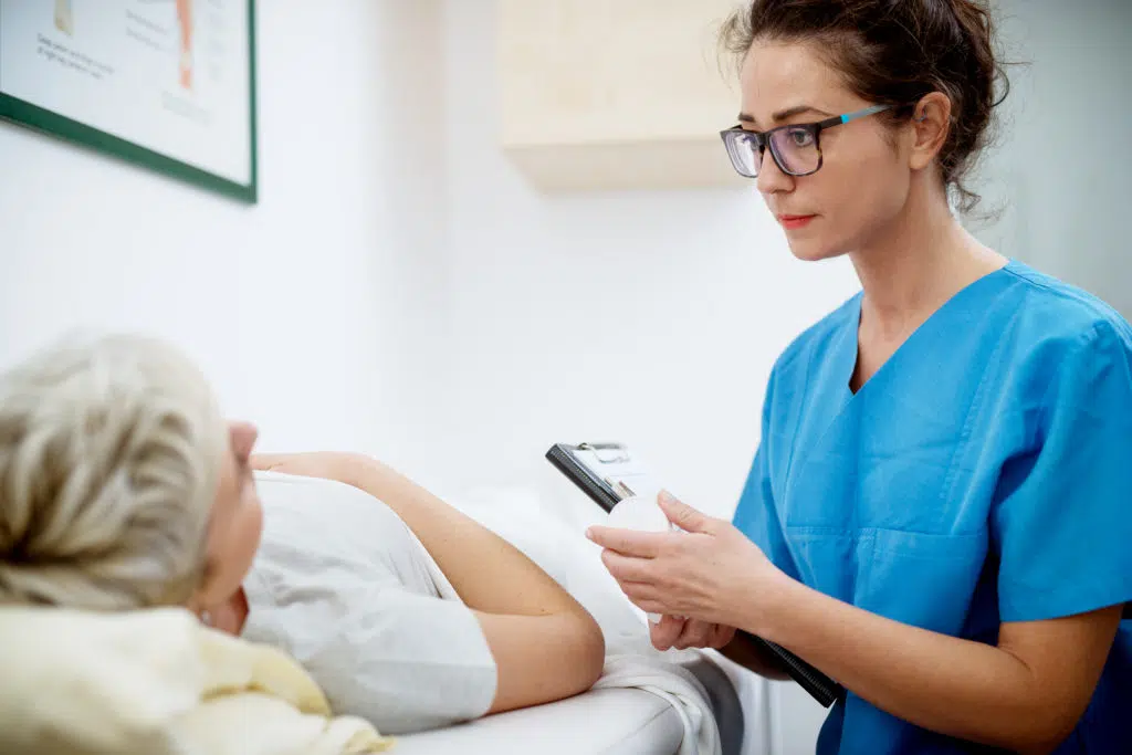
There are many benefits to participating in a clinical trial, such as the potential to access a new treatment and helping to further research into breast cancer. Another lesser-known benefit is that those who participate in a clinical trial often get more time with their treatment team.

“I have experienced quite a few benefits from being involved with the study, I had more one on one discussions with my surgeon, so I kind of felt like I had an extra layer of care, an extra layer of oversight by being involved with the study.”

“So not just being a patient but also being a participant, I feel like I got a little bit of extra care and I felt more involved with my treatment plan because I knew why I was doing what I was doing.”

“Research doesn’t always have direct benefits to the participant, but what you are almost always guaranteed is to have an involvement in contributing to the advancement of knowledge in the area and I think that’s really valuable and actually quite a cool thing to be able to say that you’ve been a part of.”

“The only way that we’re going to find better treatments and more effective treatments is by doing research, and the only way that we can do research is by people participating by donating, you know, their bodies and their time or by donating money if that’s an option for them.”

The Importance Of A Supportive Workplace and Hobbies

Laura continued to work throughout her treatment but said it wouldn’t have been possible if her workplaces were not as supportive as they were.

“Since my diagnosis, I have had to take some leave from work firstly after my initial surgery, then when I was going through chemo, I took the first week of chemo off when I knew I wasn’t going to be feeling very well.”

“The week following that, I was lucky enough that I was able to work from home, so I didn’t need to worry about the commute, and I didn’t need to worry about people being unwell near me when I was at a lower immunity.”

“And then in the third week, because I was having chemo every three weeks, when I was feeling a little better, I had the option of going into work if I did feel up to it, or just continuing to work from home or taking more sick leave if I if I needed to.”

“So, I was lucky enough to be pretty well supported by my work and by the girls in my team.”

Unfortunately for Laura, running is one of her biggest hobbies for both her physical and mental health. However due to her treatment, she hasn’t been able to “get out there and hit the pavement”.

“It just didn’t feel right for me at this stage, and I just wanted to listen to my body to see what would work best for me and running, wasn’t it unfortunately.”

“That was kind of tough that I wasn’t able to do the thing that helps me to deal with this when I was going through something like this, but it’s fine and I’m looking forward to getting back into exercise now that I’m finished active treatment.”

“I was, yeah, shocked and confused, it took a while to sink in but once I once I had kind of processed what the results were, I worked towards getting my treatment plan and once I had my treatment plan in place, that was when I started to feel better about everything because I could just see what was in front of me.”

Laura works as professional researcher at the Florey Institute of Neuroscience and Mental Health, and understands how valuable clinical trials can be.

“I am really passionate about research and I think it’s the only way that we can advance our knowledge in the area.”

“The Breast MRI Study was introduced to me and I really liked the sound of it because the idea is that at the moment the government are funding Breast MRIs for patients whose clinical examination doesn’t match what the mammogram says.”

“So for me as a younger woman, my lump didn’t show up on my on the mammogram so it was important to be able to have that extra layer of imaging with the MRI to create my treatment plan to see what was going on in there.”

“But this the funding from the government for breast MRI is limited and the government want to know well why are we doing this? Is this helpful? What are we finding from it?”

“The only way we know that is to do research and find out how it is affecting people’s treatment plans, to know if they should continue to fund it and for more people.”

“While you might not have the direct benefit to yourself, you know that you are going to be helping people down the track help other people in similar situations to yourself and their family members in there, and I think it’s important to contribute to breast cancer research either by being a participant if you are going through that yourself or financially if you are able to donate.”

Laura’s Hope For The Future

Laura participated in a clinical trial for herself and for the greater good of all breast cancer patients.

She said that although research doesn’t always have direct benefits to the participant, you are almost always guaranteed to contribute to the advancement of knowledge in that area.

“I definitely would recommend to other people to participate in clinical trials, it’s a way that you can have the extra contact with your doctor to be able to help other people in the future who are going through this”, said Laura.

“Unfortunately, research does take money to complete, so for my study it does, you know, MRIs aren’t cheap, and we need funding for participants to be able to have these MRIs.”

GIVE TO RESEARCH HELPING YOUNG WOMEN LIKE LAURA

Latest Articles

beyond the plate: rebuilding healthy habits after breast cancer
managing cording after breast cancer

Managing Cording After Breast Cancer

split-banner-image

THE FUTURE OF GENETIC RESEARCH IN BREAST CANCER

Professor Geoff Lindeman discusses genetics and breast cancer, as well as world first research in the BRCA-P clinical trial.

The Human Genome Project is an incredible medical discovery, essentially giving us the ability, for the first time, to read nature’s complete genetic blueprint for building a human being. It has revolutionised every area of human disease including cancer.

Professor Geoff Lindeman of The Walter and Eliza Hall Institute (‘WEHI’) and the Study Chair of Breast Cancer Trials BRCA-P clinical trial says, “All cancers are ultimately genetic. They’re made up of mutations that promote the growth of tumour cells. The DNA essentially goes awry.”

We now have a suite of genes that have been identified as having a link to cancer including the BRCA1 and BRCA2 genes. The identification of the BRCA genes was the culmination of a furious international hunt, call it a ‘medical space race,’ with the BRCA1 gene being discovered in 1994 and the BRCA2 gene found shortly afterwards in 1995.

The BRCA genes were identified by looking for genetic mistakes (‘mutations’) in DNA from families that had large clusters of breast and ovarian cancer to find a faulty gene. With further research it has become clear that mutations in the BRCA2 gene can also moderately increase the risk of prostate, pancreatic and even the skin cancer melanoma.

Other genes that have been linked to familial breast (and other) cancer include PALB2, TP53, PTEN, CDH1 and STK11, although the frequency of mutations in the population for some of these genes is very low.  While hereditary TP53 mutations are very rare, the TP53 gene itself is often mutated in breast and other cancers. Prof Lindeman said that some of the lessons learnt in studying familial cancers have proven helpful for understanding cancer more broadly.

Heritable mutations in other genes such as CHEK2 and ATM may also elevate breast cancer risk, but to a lesser extent than the BRCA1 genes. Knowing about mutations in these ‘moderate risk’ genes can be useful, as it helps doctors to tailor advice on breast cancer screening.

The BRCA and other genes described above are known as tumour suppressor genes. This is because they play important roles in preventing cancer by fixing mistakes in DNA that arise during normal cell division that could otherwise lead to cancer.  If a BRCA mutation is present, they are less effective at suppressing cancer. This is why cancers may occur more frequently, and often at an earlier age, compared to ‘sporadic’ forms of cancer.

There has been considerable progress in the BRCA field since the BRCA1 and BRCA2 genes were discovered just over 25 years ago. There is now a prevention trial ‘BRCA-P’ underway for BRCA1 mutation carriers. In the BRCA-P trial, Professor Geoff Lindeman and his team are investigating whether a drug called denosumab could prove a safe and effective way of preventing breast cancer in women with a faulty BRCA1 gene.

The BRCA-P study has arisen from years of work to identify the culprit cell in breast tissue that leads to breast cancer in BRCA1 mutation carriers. It turns out that the precancerous cell is activated by a signalling pathway called ‘RANK’. The drug denosumab, which is already used in the clinic to treat bone thinning (osteoporosis), works by switching off RANK signalling. The research team hopes that denosumab will also be effective at also switching off the precancerous cells in BRCA1 mutation carriers, preventing cancers from developing in the first place. “We are hopeful that denosumab, whose safety profile is well understood, can be repurposed as a prevention drug for women at the highest risk of developing breast cancer.”

Some women with a faulty BRCA1 gene undergo preventive mastectomy, which has been shown to be highly effective at preventing breast cancer. “Most women, however, don’t elect to undergo preventive mastectomy. If the study is successful, denosumab could represent a way for buying women time to delay the decision for mastectomy, or even remove the need for some women.”

Taking a pre-emptive strike at cancer is where Professor Lindeman sees the future of cancer research for women like BRCA1 mutation carriers, who have a strong hereditary predisposition. Professor Lindeman explains that “by identifying the cancer-causing genes and understanding the earliest molecular and cellular events that occur in breast tissue, it should be possible to identify ways of correcting the ‘faulty wiring’, even before cancers arise.”

“The transfer of research in this area has been quite remarkable. In the late 90’s, testing was cumbersome and slow, and focussed on families where there were dramatic clusters of breast and ovarian cancer. Nowadays people affected by cancer can undergo fairly rapid testing for a panel of genes to identify a heritable predisposition. This knowledge is being applied to guide their own management, prevent future cancers, and of course to test family members to see if they are at risk for developing breast or ovarian cancer, so that appropriate surveillance and management plans can be put in place if a mutation is found.”

The BRCA-P trial is the first international trial that hopes to proactively prevent breast cancer in BRCA1 mutation carriers. “If we can delay or prevent cancer from happening in the first place that would be a fantastic outcome. Women who take part in the study could benefit and will be contributing to a study that may benefit the next generation women” concluded Lindeman.

For more information about the BRCA-P trial go to: www.breastolution.com.au

HELP FUND WORLD-LEADING BREAST CANCER RESEARCH

Latest Articles

beyond the plate: rebuilding healthy habits after breast cancer
managing cording after breast cancer

Managing Cording After Breast Cancer

split-banner-image

PAGET’S DISEASE OF THE NIPPLE

Paget’s disease of the nipple, also known as Paget’s disease of the breast, is a rare form of breast cancer that affects the nipple and the area around the nipple (the areola). It is commonly associated with an invasive cancer elsewhere in the breast. The main sign of Paget’s disease of the nipple is a change in the nipple and/or areola.

How Common is Paget’s Disease of the Nipple?

Paget’s disease of the nipple is a rare form of breast cancer which accounts for around two of every 100 cases (2%) of breast cancer.

What are the symptoms of Paget’s Disease of the Nipple?

As the name suggests, the symptoms of Paget’s disease of the nipple usually involve a change in the nipple and/or areola. Paget’s disease of the nipple always starts in the nipple and may extend to the areola.

Symptoms of Paget’s disease of the nipple may include:

  • Itching, tingling or redness in the nipple and/or areola
  • Flaking crusting or thickened skin on or around the nipple
  • A flattened nipple
  • Yellowish or bloody discharge from the nipple.

The symptoms of Paget’s disease of the nipple may be mistaken for some benign skin conditions such as dermatitis or eczema. Due to the similarities with these conditions, some early symptoms may be misdiagnosed at first.

If you notice any changes in the skin of your nipple or areola, you should consult your GP or doctor.

How is Paget’s Disease of the Nipple Diagnosed?

A nipple biopsy will be used to correctly diagnose Paget’s disease of the nipple. A breast surgeon will perform a biopsy to remove a small piece of tissue from the nipple and/or areola area to examine it.

However, as some people with Paget’s disease of the nipple will also have an underlying breast cancer, your doctor will also do a physical exam, and will request imaging tests such as a mammogram or breast MRI.

How is Paget’s Disease of the Nipple treated?

Treatment for Paget’s disease of the nipple will depend on how much of the nipple, areola and breast is affected.

Breast surgery is the primary treatment for Paget’s disease of the nipple. This may involve a full mastectomy if the disease appears to involve a large portion of the breast, or the removal of the nipple and areola with the breast tissue underneath, known as breast conserving surgery, if it is not extensive.

Radiotherapy is commonly recommended after breast conserving surgery and is sometimes recommended after a mastectomy for patients diagnosed with Paget’s disease of the nipple.

If the invasive cancer has spread beyond the nipple, treatment with chemotherapy, radiotherapy and/or targeted or hormone therapy may be recommended.

What Are My Chances of Survival (Prognosis) if I am Diagnosed with Paget’s Disease of the Nipple?

If you are diagnosed with Paget’s disease of the breast, but it has not spread further to the surrounding breast tissue, the prognosis is excellent.

If there is an underlying cancer in the breast, survival rates decline as the stage of the cancer increases. Your age, stage of disease and tumour type has an impact on your chance of surviving five years past your breast cancer diagnosis. Those diagnosed with stage one breast cancer have an almost 100% chance of surviving five years post diagnosis, however those diagnosed with stage four breast cancer only have a 32% chance of surviving five years post diagnosis.

Overall, the five-year survival rate for women diagnosed with breast cancer is 91.5%, and 86.4% for men.

Support Us

Help us to change lives through breast cancer clinical trials research

Latest Articles

beyond the plate: rebuilding healthy habits after breast cancer
managing cording after breast cancer

Managing Cording After Breast Cancer

split-banner-image

METASTATIC BREAST CANCER

Metastatic breast cancer, also known as advanced, secondary, or stage four breast cancer, is a breast cancer which has spread to other parts of the body such as the bones, liver, or lungs. Many people who are diagnosed with metastatic breast cancer have been diagnosed with breast cancer before. However, for some it may be the first diagnosis of breast cancer.

How Does Breast Cancer Metastasise?

Metastatic breast cancer occurs when cancer cells break away from the cancer in the breast and move through the blood vessels or lymphatic vessels and form a new cancer growth in other parts of the body.

If breast cancer has spread and metastasised, it is still considered breast cancer and will be treated as such. This is because the cells which have spread are breast cancer cells. For example, if breast cancer has spread to the liver, the metastatic tumour in the liver is made up of breast cancer cells, not liver cells.

What Are the Symptoms of Metastatic Breast Cancer?

Metastatic breast cancer most commonly appears in the liver, brain, bones, or lungs (but can also occur in other parts of the body). Depending on where the cancer has spread, the following symptoms may be present. However, metastatic breast cancer can present in many different ways and if you have metastatic breast cancer, you may not present with any symptoms.

It is also important to note that some of these symptoms may not be due to metastatic breast cancer.

Bone metastasis: If breast cancer has spread to the bones, the most common symptom is a new pain or ache in the bone. Breast cancer can spread to any bone but is most commonly found in the ribs, spine, pelvis, arms, or legs.

Brain metastasis: If breast cancer has spread to the brain, you may have headaches, nausea, vomiting, vision or speech changes or memory problems. In some rare cases, symptoms can include seizures, confusion, or a change in personality.

Liver metastasis: If breast cancer has spread to the liver, symptoms may include weight loss, tiredness, and discomfort on the right side of the abdomen or stomach where the liver is located. Other less common symptoms include nausea, loss of appetite, jaundice and swelling of the abdomen.

Lung metastasis: Most commonly, the first symptom that breast cancer has spread to the lungs is a shortness of breath or a dry cough. Other less common symptoms include chest pain or a feeling of heaviness in the chest. However, it is common for breast cancer which has spread to the lung or lungs to present with no symptoms.

If you notice any of these symptoms, it is important not to panic as this may not mean your breast cancer has spread.

Consult with your doctor if you have any concerns regarding your health.

How is Metastatic Breast Cancer Diagnosed?

If your doctor suspects your breast cancer has metastasised, they will organise specific tests dependent on where they believe the cancer has spread.

To diagnose bone metastases: Bone scan, Xray, CT scan, MRI, PET scan and/or blood test.

To diagnose lung metastases: Examination of mucus under microscope, bronchoscopy, lung needle biopsy and/or surgery.

To diagnose brain metastases: MRI – often with contrast solution and a biopsy may be necessary in rare occasions.

To diagnose liver metastases: Liver function tests, MRI, CT scan, ultrasound, PET scan and/or biopsy.

How Is Metastatic Breast Cancer Treated?

Every metastatic breast cancer diagnosis is different and will therefore require different treatments. Despite the cancer growths being in other organs, such as the lung, it is called ‘breast cancer’ and is treated as breast cancer.

The aim of treating metastatic breast cancer is to control the growth and spread of the cancer, to relieve symptoms and improve or maintain quality of life.

Treatment options will depend on what is most likely to control the cancer and what side effects the patient can cope with. Treatment for metastatic breast cancer can include hormonal therapy, chemotherapy, targeted therapy, radiotherapy, and surgery.

Why Is Metastatic Breast Cancer Difficult to Treat?

There are a number of reasons why metastatic breast cancer is difficult to treat.

One reason is that many who are diagnosed with metastatic breast cancer have already been exposed to therapeutic drug treatments and the cancer has therefore have already had an opportunity to acquire some resistance. Another reason is that there is less of an opportunity to remove the cancer surgically, as the cancer has spread and become larger than it was in the primary site. Surgery is typically not used for metastatic breast cancer, apart from highly selected cases. There may also be additional genetic events that have occurred during the course the disease which, over the period of time while the cancer is regrowing, have made them more resistant to therapies.

Researchers are working to better understand why breast cancer metastasises, so they can create new and better targeted treatments.

Are There Different Kinds of Metastatic Breast Cancer?

Yes. Metastatic breast cancer means the disease has spread from the original breast cancer site located in the breast. It means it has spread to other organs in the body, most commonly the bones, liver, lungs, or brain. Metastatic breast cancer can be one of four different molecular subtypes; Luminal A (Hormone Receptor Positive HER2 Negative (HR+/HER2-) Breast Cancer), Luminal B (High grade, Hormone Receptor Positive, HER2 positive or negative (HR+/HER2+) Breast Cancer), HER2 positive breast cancer or triple negative breast cancer. The subtype of breast cancer, and the location it has metastasised will determine how the cancer is treated.

How Common is Metastatic Breast Cancer?

According to the latest data from the Australian Institute of Health and Welfare, approximately 4.6% of breast cancers diagnosed each year in Australia are stage 4. New Zealand’s incidence rates are similar.

What Are My Chances of Survival (Prognosis) If I Am Diagnosed with Metastatic Breast Cancer?

Breast cancer survival is measured in 5-year relative survival. This means how many people diagnosed with breast cancer are still alive five years after their initial diagnosis.

According to the latest data from the Australian Institute of Health and Welfare, the five-year relative survival for those diagnosed with stage 4 breast cancer is 32%. The survival rates in New Zealand are similar. This means 32% of people diagnosed with stage 4 breast cancer are alive 5 years after their diagnosis.

However, these statistics can’t predict your personal breast cancer prognosis. Breast cancer survival differs and there are many factors that can influence this such as your response to treatment, the type of breast cancer you have, medical history, overall health, age, and tumour growth. You should discuss your personal situation with your doctor and/or treatment team.

Is Metastatic Breast Cancer ‘Curable’?

Currently there is no cure for metastatic breast cancer. However new and better treatment options mean that the cancer can remain under control for longer, sometimes for years at time.

Those diagnosed with metastatic breast cancer will need to undergo treatment for the rest of their lives. If one treatment ceases to be effective in keeping the cancer under control, another treatment regime may be suggested. These treatments are generally given for as long as they are providing a benefit to the patient. The goal is to maintain the best quality of life achievable, and to prolong life if possible.

Every diagnosis of metastatic breast cancer is different, and therefore each treatment regime and prognosis will be different. Your doctor and/or treatment team are best to advise on your personal medical situation.

Support Us

Help us to change lives through breast cancer clinical trials research

Latest Articles

are we curing her+ metastatic breast cancer?

Are We Curing HER+ Metastatic Breast Cancer?

does detecting metastatic disease early make a difference?
making metastatic breast cancer manageable

Making Metastatic Breast Cancer Manageable

professor thompson on metastatic breast cancer

Professor Thompson on Metastatic Breast Cancer

split-banner-image

LUMINAL B BREAST CANCER

Luminal B breast cancer is positive for oestrogen receptor and/or progesterone receptor, is either HER2 (human epidermal growth factor receptor 2) positive or negative and is a faster growing type of breast cancer, as measured by high grade or high levels of a protein called Ki-67. Luminal B cancers tend to grow faster than the Luminal A type.

How Common is Luminal B Breast Cancer?

Around 10-20 percent of breast cancers are Luminal B tumours. Women with luminal B tumours tend to be diagnosed younger than those with Luminal A tumours.

Luminal B breast cancers are more commonly described by their hormonal status, for example, ER positive/HER2 negative and/or PR positive/HER2 negative. The luminal B molecular subtype classification is based on a special laboratory test of the tumour and is more often used in research circumstances than in discussions with patients.

2 | 1

What are the Symptoms of Luminal B Breast Cancer?

Some patients diagnosed with Luminal B may not present with any symptoms and are diagnosed on screening mammogram.

When symptoms are present, they may include:

  • A new lump in the breast, armpit area or around the collarbone
  • Thickening or hardening in the breast
  • A change in breast size or shape
  • Changes to the nipple, such as sores or crusting, an ulcer or inverted nipple
  • Clear or bloody nipple discharge
  • Changes to the skin including redness, puckering, or dimpling (an ‘orange peel’ appearance)
  • Breast tenderness or pain

If the cancer has progressed to the metastatic stage, further symptoms may be present dependent on where the cancer has spread. Learn more about metastatic breast cancer here.

Sign up to receive our latest Research Newsletter

Name(Required)

How is Luminal B Breast Cancer Diagnosed?

Luminal B breast cancer will be diagnosed in the same way as most other breast cancers. This includes a mammogram or other imaging tests. A biopsy will most likely be ordered to determine which subtype of breast cancer is present. This will help the treatment team plan treatment for the cancer.

If the breast cancer has spread and metastasised, further tests may be required to determine the size and location of the metastatic breast cancer.

How is Luminal B Breast Cancer Treated?

As Luminal B cancers are hormone receptor positive, patients with early-stage breast cancer who undergo breast surgery may receive hormonal treatments to block hormones from fuelling cancer growth.

These can include drugs such as tamoxifen, anastrozole or letrozole which are to be taken daily for at least five years in the case of early-stage breast cancer, after all other breast cancer treatments are completed.

Other treatments can include surgery, chemotherapy and/or radiotherapy. In the case of metastatic breast cancer, hormonal treatments are also used and continue for as long as they are providing a benefit to the patient.

If the cancer is HER2 positive, treatment will also likely include a HER2 targeted drug such as trastuzumab (Herceptin). The most commonly used HER2 targeted therapy is trastuzumab, sometimes in combination with pertuzumab.

This was proven to help reduce the risk of breast cancer recurrence in the early-stage breast cancer by 46%, in the Breast Cancer Trials HERA clinical trial. If the cancer is metastatic, other targeted therapies including pertuzumab (Perjeta), T-DM1 (Kadcyla) or lapatinib (Tykerb) may be used.

Learn more about Dawn’s breast cancer experience as a HERA trial participant, and how the participation of thousands of women in this research has saved lives.

What are my chances of survival (prognosis) if I am diagnosed with Luminal B Breast Cancer?

Patients with Luminal B breast cancer tend to have good survival rates, however not as high as Luminal A breast cancers. This is because Luminal B breast cancer is more aggressive and may grow and spread faster than a Luminal A breast cancer.

However, your age, stage of disease and tumour type has the greatest impact on your chance of survival. Those diagnosed with stage one breast cancer have an almost 100% chance of surviving five years post diagnosis, however those diagnosed with stage four (metastatic) breast cancer only have a 32% chance of surviving five years post diagnosis.

3 | 2

4 | 3

Overall, the five-year survival rate for women diagnosed with breast cancer is 91.5%, and 86.4% for men. Click here to find out more about male breast cancer.

Support Us

Help us to change lives through breast cancer clinical trials research

Latest Articles

beyond the plate: rebuilding healthy habits after breast cancer
managing cording after breast cancer

Managing Cording After Breast Cancer

split-banner-image

LUMINAL A BREAST CANCER?

Luminal A breast cancer is positive for oestrogen receptor and/or progesterone receptor and negative for HER2, which stands for human epidermal growth factor receptor 2. These are all growth signals that may be present on breast cancer cells. HER2 is a protein normally produced by the body. If a patient is HER2 negative it means they have a normal amount of HER2 protein.

How Common is Luminal A Breast Cancer?

Luminal A breast cancer is the most common subtype of breast cancer. It accounts for around 30-45% of all breast cancers diagnosed. Luminal A breast cancers are more commonly described as hormone receptor positive/HER2 negative, ER positive/HER2 negative, PR positive/HER2 negative or ER positive/PR positive/HER2 negative. The luminal A molecular subtype classification requires a special test to be done on a sample of the cancer and is more often used in research circumstances than in discussions with patients.

What are the Symptoms of Luminal A Breast Cancer?

Some patients diagnosed with Luminal A may not present with any symptoms and are diagnosed on screening mammogram. When symptoms are present, they may include:

  • A new lump in the breast, armpit area or around the collarbone
  • Thickening or hardening in the breast
  • A change in breast size or shape
  • Changes to the nipple, such as sores or crusting, an ulcer or inverted nipple
  • Clear or bloody nipple discharge
  • Changes to the skin including redness, puckering, or dimpling (an ‘orange peel’ appearance)
  • Breast tenderness or pain

If the cancer has progressed to the metastatic stage, further symptoms may be present depending on where the cancer has spread. Learn more about metastatic breast cancer here.

How is Luminal A Breast Cancer Diagnosed?

Luminal A breast cancer will be diagnosed in the same way as most other breast cancers. This includes a mammogram or other imaging tests. A biopsy will most likely be ordered to determine which subtype of breast cancer is present. This will help the treatment team plan treatment for the cancer.

If the breast cancer has spread and metastasised, further tests may be required to determine the size and location of the metastatic breast cancer.

How is Luminal A Breast Cancer Treated?

As Luminal A cancers are hormone receptor positive, patients with early-stage breast cancer who undergo breast surgery may receive hormonal treatments to block hormones from fuelling cancer growth. These can include drugs such as tamoxifen, anastrozole or letrozole which are taken daily for at least five years after all other breast cancer treatments are completed. These treatments can include surgery, chemotherapy and/or radiotherapy. Luminal A breast cancer may also present as metastatic, and hormone blocking medications are also used in that situation, often along with other medications to control the cancer for as long as possible. In the case of metastatic cancer, the medications continue for as long as they are effective and tolerable.

What are my chances of survival (prognosis) if I am diagnosed with Luminal A Breast Cancer?

Luminal A tumours grow at a slower rate than other cancer types, which means this subtype has a better prognosis.

However, your age, stage of disease and tumour type has the greatest impact on your chance of survival. Those diagnosed with stage one breast cancer have an almost 100% chance of surviving five years post diagnosis, however those diagnosed with stage four Luminal A breast cancer have a 50% chance of surviving five years post diagnosis.

Overall, the five-year survival rate for women diagnosed with breast cancer is 91.5%, and 86.4% for men.

Support Us

Help us to change lives through breast cancer clinical trials research

Latest Articles

beyond the plate: rebuilding healthy habits after breast cancer
managing cording after breast cancer

Managing Cording After Breast Cancer

split-banner-image

LOCALLY ADVANCED BREAST CANCER?

Locally advanced breast cancer is an invasive breast cancer that is large (typically 5cm or more) or has spread to areas near the breast, such as the chest wall. However, there are no signs the cancer has spread beyond the breast region or to other parts of the body. It also called stage 3 breast cancer.

What is Locally Advanced Breast Cancer?

Locally advanced breast cancer is an invasive breast cancer that is large (typically 5cm or more) or has spread to areas near the breast, such as the chest wall. However, there are no signs the cancer has spread beyond the breast region or to other parts of the body. It also called stage 3 breast cancer.

Stage 3 breast cancer can be stage 3 A, stage 3 B, or stage 3 C:

  • Stage 3A – The tumour is less than 5cm and has spread to 4-9 lymph nodes in armpit or under breastbone. Or the tumour is more than 5cm and has spread to 1-9 lymph nodes.
  • Stage 3B – The cancer has spread to nearby muscles and skin. The tumour can be any size.
  • Stage 3C – The cancer has spread to at least 10 lymph nodes in armpit, or at least one node under breastbone and at least one node in the armpit, or to at least one node near collarbone. The tumour can be any size.

What are the Symptoms of Locally Advanced Breast Cancer?

Some patients diagnosed with locally advanced breast cancer may not present with any symptoms. When symptoms are present, they may include:

  • A lump in the breast or armpit that doesn’t move freely but feels attached to the chest wall
  • A lump at the base of the neck
  • A red and/or swollen breast (this is called inflammatory breast cancer)
  • Ulcers on the breast
  • Thickening or hardening in the breast
  • A change in breast size or shape
  • Changes to the nipple, such as sores or crusting, an ulcer or inverted nipple
  • Clear or bloody nipple discharge
  • Changes to the skin including redness, puckering, or dimpling (an ‘orange peel’ appearance)
  • Breast tenderness or pain

How is Locally Advanced Breast Cancer Diagnosed?

Locally Advanced breast cancers breast cancers may be large enough that it may be felt as a hard lump during a physical examination, or self-examination. However, these breast cancers may also be diagnosed through screening mammograms. An ultrasound may be necessary to further investigate the spread of disease.

A biopsy may be conducted to learn more about the tumour/s before treatment.

A CT, bone scan are usually also ordered (and in some circumstances a PET scan) to check that the cancer has not spread to other parts of the body.

How Common is Locally Advanced Breast Cancer?

According to the latest data from the Australian Institute of Health and Welfare, approximately 12% of breast cancers diagnosed each year in Australia are stage 3. New Zealand’s incidence rates are similar. Thanks to the successful BreastScreen Australia and BreastScreen Aotearoa programs, more breast cancers are being diagnosed in the earlier stages which is leading to higher survival rates.

Are there different kinds of Locally Advanced Breast Cancer?

Yes. Locally advanced breast cancer can be one of four different molecular subtypes; Luminal A (Hormone Receptor Positive, HER2 Negative (HR+/HER2-) low grade), Luminal B (Hormone Receptor Positive, HER2 negative/positive (HR+/HER2-/+) high grade), HER2 positive breast cancer or triple negative breast cancer. Locally advanced breast cancer can also be a rarer form of the disease called Inflammatory breast cancer.

How is Locally Advanced Breast Cancer Treated?

Treatment for locally advanced breast cancer will usually involve a combination of breast surgery, chemotherapy, radiotherapy, targeted therapies, or hormonal therapies. Commonly, chemotherapy will be given before surgery. However, treatment will differ from person to person depending on the sub-type of breast cancer they have, the extent and size of the breast cancer, the location of the breast cancer, age, general health, and the patients treatment preferences.

What are my chances of survival (prognosis) if I am diagnosed with Locally Advanced Breast Cancer?

Breast cancer survival is measured in 5-year relative survival. This means how many people diagnosed with breast cancer are still alive five years after their initial diagnosis.

According to the latest data from the Australian Institute of Health and Welfare, the five-year relative survival for those diagnosed with stage 3, or locally advanced, breast cancer is 80.6%

However, breast cancer survival differs and there are many factors that can influence this such as your response to treatment, the type of breast cancer you have, medical history, overall health, age, and tumour growth. You should discuss your personal situation with your doctor and/or treatment team.

Support Us

Help us to change lives through breast cancer clinical trials research

Latest Articles

beyond the plate: rebuilding healthy habits after breast cancer
managing cording after breast cancer

Managing Cording After Breast Cancer

split-banner-image

WHAT IS LOBULAR CARCINOMA IN SITU?

Lobular carcinoma in situ or LCIS is a non-invasive breast condition. LCIS is the name for abnormal cells that are contained to the terminal ductal lobular units (milk glands) of the breast. It is considered a breast change, and not breast cancer.

What is Lobular Carcinoma in Situ or LCIS?

Lobular carcinoma in situ or LCIS is a non-invasive breast condition. LCIS is the name for abnormal cells that are contained to the lobules (milk glands) of the breast. It is considered a breast change, and not breast cancer.

What are the Symptoms of Lobular Carcinoma in Situ?

LCIS cannot usually be felt as a breast lump and does not usually present with symptoms sometimes seen in invasive breast cancers. Changes due to LCIS are only sometimes found on a mammogram.

Most cases of LCIS are found when a patient is undergoing a breast biopsy for another reason. Most patients are not aware of any symptoms at the time of diagnosis.

How is Lobular Carcinoma in Situ Diagnosed?

LCIS cannot usually be felt in the breast and usually isn’t visible on a mammogram. LCIS is diagnosed by a biopsy, where small pieces of breast tissue are removed and checked under a microscope in the lab.

How is Lobular Carcinoma in Situ Treated?

LCIS does not require treatment if there are no other abnormal changes to the breast. This is because LCIS typically does not spread beyond the lobule and become invasive breast cancer if it isn’t treated.

However, a woman with LCIS should be carefully monitored as having LCIS increases the chance of developing breast cancer in future. If you have been diagnosed with LCIS, your doctor may recommend more frequent physical exams and screening to monitor your breasts for signs of cancer.

In some cases, breast conserving surgery or preventative medication may be recommended to reduce the risk of invasive breast cancer developing in the future.

Is Lobular Carcinoma in Situ Life-Threatening?

Like DCIS, although these abnormal changes can turn into invasive cancer cells, it is not classified as breast cancer as we generally understand it. A woman cannot die from LCIS as the cells are contained to the lobules of the breast.

Support Us

Help us to change lives through breast cancer clinical trials research

Latest Articles

beyond the plate: rebuilding healthy habits after breast cancer
managing cording after breast cancer

Managing Cording After Breast Cancer

split-banner-image

WHAT IS INVASIVE LOBULAR CARCINOMA?

Invasive lobular carcinoma (ILC) is a type of breast cancer that has spread beyond the lobules and ducts, potentially spreading to the lymph nodes and other parts of the body.

Lobular Breast Cancer

Lobular breast cancer is a subtype of breast cancer that originates in the lobules, the glands that produce milk in the breast tissue. This differs from the more common ductal carcinoma, which starts in the milk ducts. It is classified as invasive when cancer cells spread beyond the lobules into surrounding breast tissue.

The exact causes of lobular breast cancer remain unclear, but several risk factors have been identified. These include genetic mutations (such as alterations in the CDH1 gene), hormonal factors (such as increased estrogen levels), and a family history of breast cancer. One of the challenges with lobular breast cancer is that it often does not present with a distinct lump, making early detection through mammograms or breast self-examination difficult.

Early diagnosis is crucial for effective treatment and improved outcomes. Treatment options typically include a combination of surgery, radiation therapy, chemotherapy, endocrine (or hormone) therapy, and/or targeted therapies, depending on the stage and characteristics of the cancer. Ongoing advances in research continue to enhance understanding and treatment options for lobular breast cancer, offering hope for improved outcomes and quality of life for those affected.

What are the Symptoms of Invasive Lobular Carcinoma?

Some patients diagnosed with ILC may not present with any symptoms. When symptoms are present, they may include:

  • A new lump in the breast, armpit area or around the collarbone.
  • Thickening or hardening in the breast.
  • A change in breast size or shape.
  • Changes to the nipple, such as sores or crusting, an ulcer or inverted nipple.
  • Clear or bloody nipple discharge.
  • Changes to the skin including redness, puckering or dimpling (an ‘orange peel’ appearance).
  • Breast tenderness or pain.

How is Lobular Breast Cancer Diagnosed?

Diagnosing ILC can be challenging due to the way the cells grow through the breast tissue. ILC typically grows in a line rather than a mass, making it difficult to see the full extent of the disease on a mammogram. Additional imaging with an ultrasound and breast MRI is sometimes required.

Imaging Tests

  • Mammogram: A specialised X-ray of the breast to detect any abnormalities or unusual growths. However, ILC can be difficult to identify on mammograms.
  • Ultrasound: This technique uses sound waves create images, providing a clearer view, especially if the mammogram raises suspicions.
  • Breast MRI: A more detailed imaging test that can provide a comprehensive picture of the breast tissue.

Biopsy

If an imaging test shows an abnormality, a biopsy is performed. This involves taking a small sample of tissue from the suspicious area. A core needle biopsy is commonly used, where a thin, hollow needle is used to collect tissue for examination.

Pathology Examination

The collected tissue is sent to a laboratory, where a pathologist examines it under a microscope. The pathologist looks for specific characteristics, such as the presence of invasive lobular structures.

Hormone Receptor Testing

Testing for oestrogen and progesterone receptors helps determine if the cancer cells are sensitive to endocrine (or hormone) therapies.

Lymph Node Examination

If cancer is confirmed, the doctor may examine nearby lymph nodes to see if the cancer has spread. This is an important step in determining the extent or stage of the cancer.

Staging

Understanding the stage of cancer is crucial for planning the most appropriate treatment. Staging considers the size of the tumour, whether it has spread to the lymph nodes, and whether it has spread to other parts of the body.

Stages of Lobular Carcinoma

Invasive lobular carcinoma (ILC) goes through different stages. It is crucial for doctors to know the stage of the cancer to assess the severity of the disease and guide the treatment plan. The stages of cancer are:

  • Stage 0 (Carcinoma in Situ): At this early stage, cancerous cells are confined to the lobules, and there is no invasion into surrounding tissues. This is often referred to as carcinoma in situ, meaning the cancer is localised and has not spread.
  • Stage I: Cancer is still relatively localised, typically confined to the breast tissue. The tumour size is relatively small, and there is no evidence of lymph node involvement.
  • Stage II: The tumour is larger or may involve nearby lymph nodes. Despite the larger tumour size, the cancer is still predominantly within the breast.
  • Stage III: Cancer has spread more extensively to surrounding tissues and lymph nodes. The tumour may be large, and there may be evidence of invasion into nearby structures.
  • Stage IV (Metastatic): At this advanced stage, cancer has spread beyond the breast and nearby lymph nodes to other organs or distant parts of the body. This is considered advanced and challenging to treat.

How is Invasive Lobular Carcinoma (ILC) Treated?

ILC are typically oestrogen receptor positive breast cancers, which makes them sensitive to the endocrine (or hormone blocking) treatments such as tamoxifen.

Depending on the stage of ILC, treatment may include a combination of surgery, radiation therapy, chemotherapy, endocrine (hormone) therapy, and targeted therapies. Treatment for ILC also may include breast surgery, radiation therapy and chemotherapy. In most cases, ILC is usually more responsive to endocrine therapy than chemotherapy and is generally preferred.

What are my Chances of Survival (Prognosis) if I am Diagnosed with Invasive Lobular Carcinoma?

Typically, invasive lobular carcinoma tumours are associated with a good prognosis, as they tend to be low grade and oestrogen receptor positive. However, this will depend on your age, health status, tumour type and stage of cancer.

Your age, stage of disease and tumour type has the greatest impact on your chance of surviving five years past your breast cancer diagnosis. For stage 1 breast cancer, the chance of surviving 5 years or beyond is nearly 100%. However, for stage 4 breast cancer, the chance of surviving 5 years or beyond drops to 22%.

Overall, the five-year survival rate for women diagnosed with breast cancer is 91.5%, and 86.4% for men.

Support Us

Help us to change lives through breast cancer clinical trials research

Latest Articles

beyond the plate: rebuilding healthy habits after breast cancer
managing cording after breast cancer

Managing Cording After Breast Cancer

split-banner-image

WHAT IS INVASIVE DUCTAL CARCINOMA?

Invasive ductal carcinoma (IDC) is a broad term used to classify cancer that began growing in a milk duct and has invaded the fibrous or fatty tissue of the breast outside the duct.

Invasive ductal carcinoma (IDC) is a type of breast cancer that originates in a milk duct and invades the surrounding fibrous or fatty tissue of the breast. From there, it has the potential to spread to other organs in the body.

How Common is Invasive Ductal Carcinoma?

Invasive ductal carcinoma accounts for around 80 percent of all breast cancer diagnoses. It is also the most common type of breast cancer that affects men.

What are the Symptoms of Invasive Ductal Carcinoma?

Some patients diagnosed with IDC may not present with any symptoms. When symptoms are present, they may include:

  • A new lump in the breast, or armpit area
  • A change in breast size or shape
  • Changes to the nipple, such as sores or crusting, an ulcer or inverted nipple
  • Clear or bloody nipple discharge
  • Changes to the skin including redness, puckering, or dimpling (an ‘orange peel’ appearance)
  • Breast tenderness or pain

How is Invasive Ductal Carcinoma Diagnosed?

Invasive ductal carcinoma is diagnosed through a combination of a physical exam, imaging tests like mammogram and ultrasound, and a biopsy where a small tissue sample is examined for cancer cells. This help doctors confirm the presence of cancer and plan the best treatment.

How is Invasive Ductal Carcinoma Treated?

The treatment of IDC depends on the stage of the cancer and the characteristics of the tumour, including its size, location, spread, and type. Treatment options may include a combination of radiation therapy, chemotherapy, hormone therapy, and/or surgery.

What are my chances of Survival (prognosis) if I am Diagnosed with Invasive Ductal Carcinoma?

In Australia and New Zealand, survival rates for invasive ductal carcinoma have improved thanks to advancements in clinical trials research, which have led to more tailored and advanced treatments, prevention strategies, and screening techniques.

However, for some people, their breast cancer will progress to an advanced or metastatic stage which greatly reduces their chances of survival.

Factors such as age, stage of disease, and tumour type can impact the likelihood of surviving five years past a breast cancer diagnosis.

Overall, the five-year survival rate for women diagnosed with breast cancer is 91.5%, and 86.4% for men.

FAQ’s

How Serious is Invasive Ductal Carcinoma?

Invasive ductal carcinoma is a serious form of breast cancer, as it has the potential to spread beyond the milk ducts where it originates. The seriousness of IDC largely depends on the grade of the cancer and the stage at which it is diagnosed.  Early detection and appropriate treatment are crucial for a better prognosis.

Is Invasive Breast Cancer Aggressive?

Yes, invasive breast cancer, including invasive ductal carcinoma, is considered aggressive because it can invade surrounding tissues and spread to other parts of the body. The level of aggressiveness can vary depending on the grade of the cancer, with invasive ductal carcinoma   grade 3 being more aggressive compared to grade 1.

What is the Difference Between DCIS and Invasive Ductal Carcinoma?

The key difference between DCIS (ductal carcinoma in situ) and invasive ductal carcinoma lies in their potential to spread. DCIS is a non-invasive cancer, meaning the cancer cells are confined within the ducts and have not invaded surrounding tissues. On the other hand, invasive ductal carcinoma breaks through the duct walls and invades the surrounding breast tissue, which can lead to metastasis if not treated effectively.

What is the Difference Between Invasive Ductal Carcinoma and Metastatic Breast Cancer?

While invasive ductal carcinoma refers to cancer that has invaded surrounding breast tissue, metastatic breast cancer occurs when the cancer cells spread from the original tumour site to distant organs of the body, such as the bones, liver, lungs, or brain. This spread, known as metastasis, marks an advanced stage and will change the treatment plan and reduces survival rates.

What is the Invasive Ductal Carcinoma Recurrence Rate?

The recurrence rate of early invasive ductal carcinoma recurrence rate varies depending on several factors including stage of diagnosis, tumour grade, hormone receptor and HER2 receptor status as well as effectiveness of the initial treatment. Generally, higher grades and stages of invasive ductal carcinoma have higher risks of recurrence.

Recurrence can happen locally (near the site of the original tumour), regionally (nearby lymph nodes), or in distant organs. Effective initial treatment and ongoing surveillance are critical in managing the risk of recurrence and improving long-term outcomes.

Support Us

Help us to change lives through breast cancer clinical trials research

Latest Articles

beyond the plate: rebuilding healthy habits after breast cancer
managing cording after breast cancer

Managing Cording After Breast Cancer

split-banner-image

HER2-POSITIVE BREAST CANCER

HER2-positive breast cancer is a type of breast cancer that tests positive for a protein called human epidermal growth factor receptor 2 (HER2). An excess amount of HER2 promotes the growth of this type of breast cancer.

What is HER2-Positive Breast Cancer?

HER2 positive breast cancer is a type of breast cancer that tests positive for a protein called human epidermal growth factor receptor 2 (HER2). This protein promotes the growth of cancer cells.

HER2-positive breast cancer tends to grow faster compared with HER2 negative ones. However, they often respond well to treatments specifically designed for HER2-positive cancers. These cancers can either be hormone-receptor (HR) positive or negative.

How Common is HER2-Positive Breast Cancer?

Around 15-20% of all types of breast cancers are HER2-positive. It is more common in younger, pre-menopausal women.

What are the Symptoms of HER2-Positive Breast Cancer?

Some patients diagnosed with HER2-positive breast cancer may not present with any symptoms. When symptoms are present, they may include:

  • A new lump in the breast, armpit area or around the collarbone
  • Thickening or hardening in the breast
  • A change in breast size or shape
  • Changes to the nipple, such as sores or crusting, an ulcer or inverted nipple
  • Clear or bloody nipple discharge
  • Changes to the skin including redness, puckering, or dimpling (an ‘orange peel’ appearance)
  • Breast tenderness or pain

If the cancer has progressed to the metastatic stage, additional symptoms may be present depending on where the cancer has spread. Learn more about metastatic breast cancer here.

How is HER2-Positive Breast Cancer Diagnosed?

HER2-positive breast cancer is diagnosed in the same way as most other breast cancers, which typically involves a mammogram and a biopsy. The biopsy is important for determining the HER2 status of the cancer, which helps to guide the treatment plan for the treating doctors. Sometimes the HER2 test is performed on the cancer tissue once it has been surgically removed.

If the breast cancer has spread to other organs, additional tests may be needed to determine the extent and location of the spread.

How is HER2-Positive Breast Cancer Treated?

HER2-positive breast cancer is primarily treated with HER2-targeted therapies. The most common of these in Australia and New Zealand is trastuzumab (Herceptin). The HERA clinical trial found Trastuzumab significantly reduced the likelihood of early HER2-positive breast cancer returning.

Women with early HER2-positive breast cancer may also receive this in combination with surgery, chemotherapy, other HER2-targeted treatments and radiation therapy.

For women with metastatic HER2-positive breast cancer, trastuzumab may be combined with other treatments including other HER2-targeted therapies. This will continue so long as the benefit o the patient outweighs the side effects.

If the breast cancer is also Hormone Receptor-positive (HR-positive), it will grow as a result of the female hormones oestrogen and/or progesterone. Hormone blocking (or endocrine) treatments may also be prescribed in this situation.

What are my chances of Survival (prognosis) if I am Diagnosed with HER2-Positive Breast Cancer?

The prognosis for HER2-positive breast cancer prognosis has improved significantly thanks to targeted treatments like trastuzumab (Herceptin).

Factors such as age, stage of disease, and tumour type greatly impact your chance of survival. Those diagnosed with stage one breast cancer have a 96% chance of surviving five years after diagnosis, while those diagnosed with stage four HER-2 positive breast cancer have over 50% chance of surviving five years after diagnosis of metastatic disease.

Overall, the five-year survival rate for women diagnosed with breast cancer is 91.5%, and 86.4% for men.

What Clinical Trials are Available for HER2 Breast Cancer Patients?

Clinical trials have been vital in advancing the treatment of HER2-positive breast cancer. 

To understand the experience of participating in a clinical trial, discover Laura’s story.

Supporting the latest breast cancer clinical trials today is the best way to change breast cancer outcomes – for you and your loved ones – tomorrow. Donate to improve treatment and save lives.

FAQ’s

Is HER2-Positive Breast Cancer Curable?

With the advancements in treatment, HER2-positive breast cancer is increasingly curable, especially when diagnosed early. The effective targeted therapies, like trastuzumab, have significantly improved the outcomes and life expectancy for HER-2 positive breast cancer.

What Does HER2-Positive Mean?

When a breast cancer is HER2-positive, it means that the cancer cells have an excess of the HER2 gene, leading to an overproduction of the HER2 protein. This overexpression helps the cancer cells grow and divide faster, but it also makes them more susceptible to targeted HER2 therapies.

Does HER2-Positive Breast Cancer Always Require Chemotherapy?

HER2-positive breast cancer treatment often includes chemotherapy, but it is not always required. The need for chemotherapy depends on factors such as size, grade and stage of the cancer. Targeted therapies can sometimes be used in combination with or instead of chemotherapy.

Is it Better to Have HER2-Positive or Negative Breast Cancer?

It is difficult to say whether it is better to have HER2-positive or HER2-negative breast cancer as both types have different treatment approaches and prognosis. However, HER2-positive breast cancer tends to respond well to targeted treatments, which have dramatically improved outcomes for this group.

Is HER2 Breast Cancer Aggresive?

HER2 breast cancer is typically more aggressive than other types of breast cancer due to its rapid growth rate. However, this type of cancer is also more responsive to specific targeted treatments, which can lead to better outcomes than some less aggressive, harder-to-treat cancers.

Support Us

Help us to change lives through breast cancer clinical trials research

Latest Articles

beyond the plate: rebuilding healthy habits after breast cancer
managing cording after breast cancer

Managing Cording After Breast Cancer

split-banner-image

EARLY STAGE BREAST CANCER

Early breast cancer is an invasive breast cancer that is contained in the breast and may or may not have spread to the lymph nodes in the breast or armpit. If you have been diagnosed with Stage 1 or Stage 2 breast cancer, you have early-stage breast cancer.

What is Early-Stage Breast Cancer?

Early breast cancer is an invasive breast cancer that is contained in the breast and may or may not have spread to the lymph nodes in the breast or armpit. If you have been diagnosed with Stage 1 or Stage 2 breast cancer, you have early-stage breast cancer.

 Stage 1: A stage one breast cancer means the cancer cells has not spread from the original site to fatty tissues and/or lymph nodes. The tumour will be less than 2cm.

Stage 2: The cancer is larger than Stage 1 and/or has spread to nearby lymph nodes but has not spread beyond the breast tissue or lymph nodes. In stage 2 breast cancer, the tumour is less than 2cm and has spread to 1-3 lymph nodes in the armpit, or the tumour is between 2cm-5cm and has not spread to the lymph nodes.

What are the Symptoms of Early-Stage Breast Cancer?

Some patients diagnosed with early-breast cancer may not present with any symptoms. When symptoms are present, they may include:

  • A new lump in the breast, or armpit area
  • Thickening or hardening in the breast
  • A change in breast size or shape
  • Changes to the nipple, such as sores or crusting, an ulcer or inverted nipple
  • Clear or bloody nipple discharge
  • Changes to the skin including redness, puckering, or dimpling (an ‘orange peel’ appearance)
  • Breast tenderness or pain

How is Early-Stage Breast Cancer Diagnosed?

As stage 1 breast cancer is small, it may be difficult to detect in a physical examination. Often stage one breast cancers are picked up in routine screening mammograms.

Stage 2 breast cancers are slightly larger, with the tumour measuring between 2cm to 5cm, and so it may be felt as a hard lump in the breast or under the arm during a physical examination, or self-examination. Stage two breast cancers are also often diagnosed through screening mammograms.

If early-stage breast cancer is suspected, your doctor may also request you have other scans such as an ultrasound or MRI. A biopsy of the tumour may also be needed to confirm the diagnosis and determine what type of breast cancer you have, which will help to influence your treatment regimen.

How Common is Early-Stage Breast Cancer?

According to the latest data from the Australian Institute of Health and Welfare, approximately 42% of breast cancers diagnosed each year in Australia are stage 1, and 35% are stage 2. New Zealand’s incidence rates are similar. Thanks to the successful BreastScreen Australia and BreastScreen Aotearoa programs, more breast cancers are being diagnosed in the earlier stages which is leading to higher survival rates.

Are there different kinds of Early-Stage Breast Cancer?

Yes. Early-stage breast cancer means that the breast cancer has not spread beyond the breast or nearby lymph nodes. Early-stage breast cancer can be one of four different major subtypes; Luminal A (Hormone Receptor positive, HER2 negative (HR+/HER2-) low grade), Luminal B (Hormone Receptor positive, HER2 negative or positive (HR+/HER2-/+) high grade), HER2 positive breast cancer or triple negative breast cancer.

Your treatment will often depend on what subtype of breast cancer you have.

How is Early-Stage Breast Cancer Treated?

The aim of treatment for early breast cancer is to remove the breast cancer and any cancer cells that may be in the breast or armpit. Treatment may also be needed for cancer cells that may have spread to other parts of the body but cannot be detected.

Treatment can involve radiotherapy, breast surgery, chemotherapy, hormonal therapies, and targeted therapies.

What are my chances of survival (prognosis) if I am diagnosed with Early-Stage Breast Cancer?

Breast cancer survival is sometimes measured in 5-year relative survival. This means how many people diagnosed with breast cancer are still alive five years after their initial diagnosis. Many of those who are alive at 5 years will still be alive and cancer-free for many years after that point.

Breast cancer survival differs and there are many factors that can influence this such as your response to treatment, the type of breast cancer you have, medical history, overall health, age, and tumour growth. You should discuss your personal situation with your doctor and/or treatment team.

Support Us

Help us to change lives through breast cancer clinical trials research

Latest Articles

beyond the plate: rebuilding healthy habits after breast cancer
managing cording after breast cancer

Managing Cording After Breast Cancer