EVALUATING THE CLINICAL PERFORMANCE OF NASHA GEL
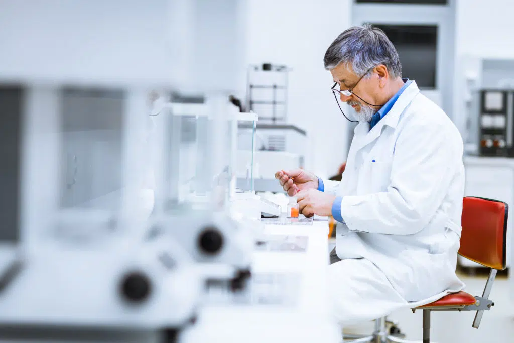
As part of a Clinical Fellowship project with Breast Cancer Trials, Dr Janice Yeh is evaluating the technical feasibility and clinical performance of the novel NASHA Gel compared to standard surgical clips as a fiducial marker.
Evaluating the Clinical Performance of NASHA Gel Compared to Surgical Clips
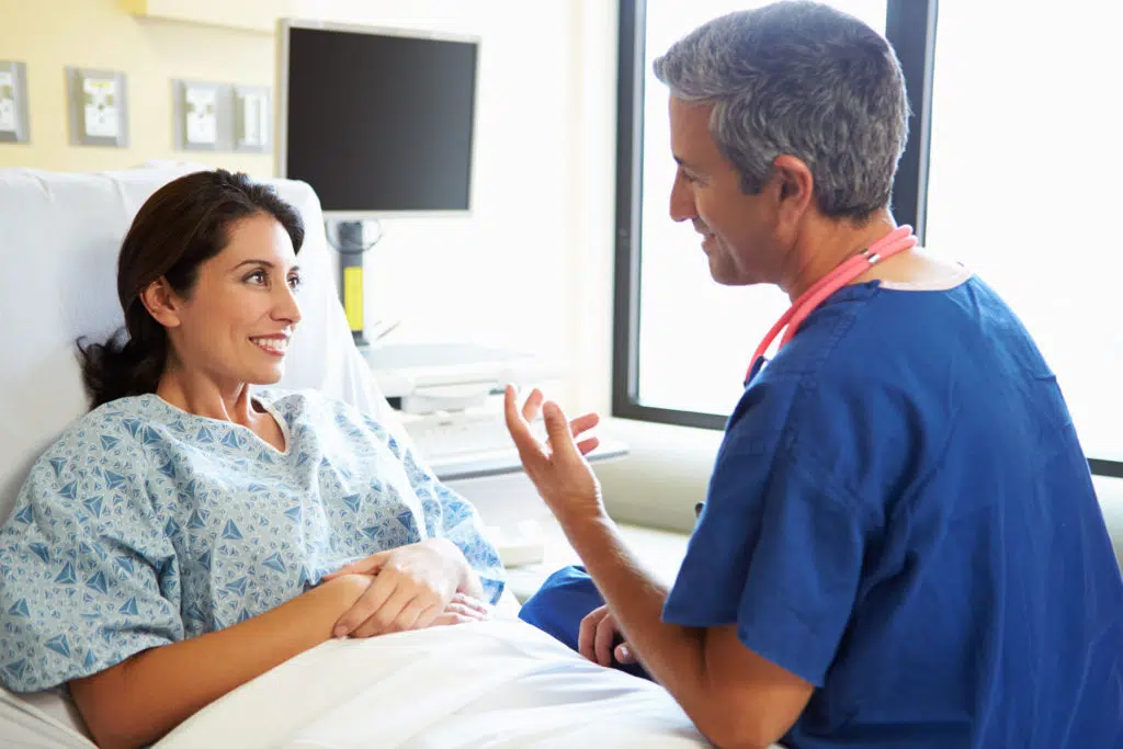
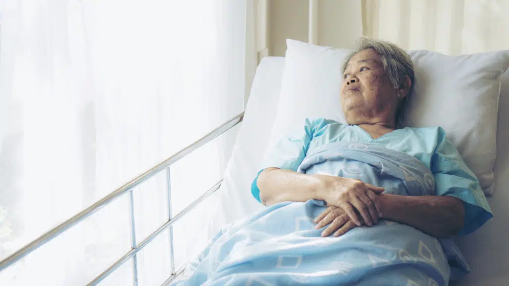
Radiation therapy after breast conserving surgery plays an important role in the management of early breast cancer, decreasing the risk of breast cancer returning and improving mortality rates.
As part of a Clinical Fellowship project with Breast Cancer Trials, Dr Janice Yeh is evaluating the technical feasibility and clinical performance of the novel NASHA Gel compared to standard surgical clips as a fiducial marker. We asked Dr Yeh to explain the role that radiation therapy has in the treatment of early breast cancer.
“So, radiotherapy targeting the breast tissue after a lumpectomy surgery for early breast cancer improves long term survival outcomes and local control. It’s basically the equivalent to having a mastectomy, which is having the breast completely surgically removed.”
“It allows women to preserve their breast tissue without compromising on oncological outcomes.”
Listen to the podcast
Listen to our conversation with Clinical Fellow, Dr Janice Yeh, as she discusses her research project which is evaluating the technical feasibility and clinical performance of the novel NASHA Gel.
What is a Fiducial Marker, and How are Surgical Clips Presently Used?
“Fiducial markers in general are usually small metallic objects that are placed near, or at the site where we want to treat, and it helps us be more accurate with targeting the treatment.”
“Surgical clips have been around for many years. They do stay in the body usually permanently, however you shouldn’t be able to feel it. The potential downside to them is that we are limited in the number of clips we can put in because, they can potentially cause metallic interference when it comes to scans that patients might have in the future.”
What is NASHA Gel?
NASHA gel is a type of hyaluronic acid, which is a common ingredient in serums and creams, and it’s a naturally occurring substance in the body. Dr Yeh explains why NASHA gel might be better than surgical clips as a fiducial marker.
“NASHA stands for ‘Non-Animal Stabilized Hyaluronic Acid’, which just means it’s a manufactured form of hyaluronic acid where it’s, stabilized in a manner that improves shelf life and longer half-life for clinical use.”
“Because of the gel like consistency of it, the surgeon is able to inject little dots of it as points of reference for the fiducial marking of the tumour bed to help with being more accurate with radiotherapy planning.”
“It’s designed to be biodegradable over time, and so we can put as many dots in as needed, we’re not restricted in the same way that we might be with clips.”
“So as an example, a common number of clips useful for fiducial marking is probably around four, whereas with our study there can be as many as 16 dots put in when using the gel. If you think about it, having more points should allow us to be more accurate and be more consistent with knowing where we’re treating, and therefore targeting the most at risk area of the breast after breast cancer surgery.”
“If we can prove that the NASHA gel allows us to be more accurate than surgical clips, then more patients may be offered partial breast radiotherapy because then there’s less of a worry that we’re missing the target.”
An Overview of Dr Yeh’s Project
Developing new research ideas and supporting the next generation of researchers is at the heart of the new Clinical Fellowship Program.
The Program is aimed at early career researchers, who have a high-level interest in clinical research and qualifications in the disciplines of medical oncology, pathology, psychology and other supportive care specialties, radiation oncology, radiology or surgery.
“So, my research with Breast Cancer Trials is a pilot feasibility study, where we are wanting to compare the consistency of being able to draw out the tumour bed using the clips versus the gel.”
“At the time of surgery after the lump is removed, the surgeon puts in both the gel and the clips into the same patient. Then when it comes time to planning the radiotherapy, we have the patient undergo a CT scan as well as an MRI scan. We then use those images visualize the fiducial markers and compare the consistency with delineating the tumour bed.”
“So, we have a team of six observers, five radiation oncologists and one radiologist, and once the patient’s scans are done, they sit down and draw out what they think the tumour bed is. We then compare against each other to see which using which fiducial results in the least inter-observer variation.”
“We started recruitment for this project last year, and as of late May 2022, we’ve recruited more than three quarters of our target number of patients.”
“It’s very exciting that we’ve had a very good engagement with patients, as well as our multidisciplinary team, and we hope to be able to publish some results in the next 12-months.”
Dr Yeh’s Hope for the Future
“I mean obviously I’d like to be able to have firmer results before we can be clearer about our next step. The company that I’m working with to produce the NASHA gel, are in development of a radio opaque version of the gel. So once that comes along, I think it would be important to also validate our results with this newer form of the gel and then make it make it more accessible.”
“We are also looking to increase follow up in terms of formally reviewing the patient’s annual post op mammograms with a specialist breast radiologist, to see if the gel is still visible. The degradation varies quite a lot depending on the amount that was injected, where it was injected, and the patient’s natural biology. So, we think it’s really important to assess this more formally.”
To find out more about Breast Cancer Trials Clinical Fellowship Program, follow the link below.